Coat Color Genetics in Border Collies and Our Approach to Healthy Breeding
Introduction — Coat Color Is More Than Aesthetics
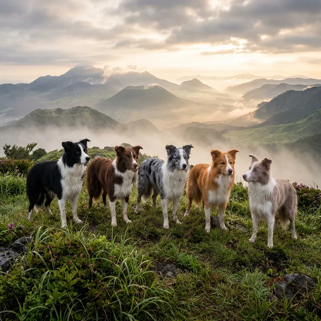
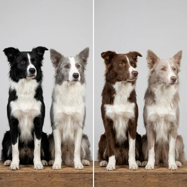
The Border Collie boasts a remarkably diverse color palette. From jet-black and white to deep chocolate, silvery blue, ethereal lilac, fiery red, and the distinctively mottled blue merle — this chromatic variety is undeniably one of the breed’s great attractions.
But coat color is far more than a matter of appearance.
Some of the genes governing coat color are directly linked to health. Double merle (M/M) individuals face serious risks of visual and auditory impairment. Color dilution alopecia (CDA) threatens the skin health of dilute-colored dogs. Excessive white markings correlate with congenital deafness. All of these are risks determined by specific combinations of coat color genes.
At ROSCH KENNEL, nestled within the landscapes of Kirishima National Park, breeding is guided by scientific evidence. We believe that beauty and health are not in opposition — both can be achieved through proper genetic knowledge and responsible management. This article offers a thorough explanation of the seven major genetic loci that determine Border Collie coat color, along with practical knowledge for sound breeding.
How Coat Color Works — Two Types of Melanin
Canine coat color is fundamentally determined by the quantity and distribution of two types of melanin pigment:
- Eumelanin — Black-to-brown pigment. Deposited not only in the coat but also in the nose leather, eyelid rims, lips, and paw pads.
- Phaeomelanin — Red-to-yellow pigment. Ranges from pale cream to deep red.
Every coat color in every dog is the result of combinations in production volume, distribution pattern, and modification (dilution/mutation) of these two pigments. The following seven genetic loci control when, where, and how much of each pigment is produced.
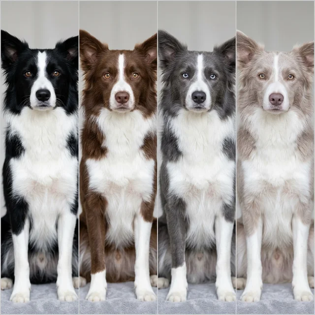
In Border Collies, eumelanin base colors are classified into four categories through genetic modification:
- Black — B/B or B/b × D/D or D/d (undiluted black)
- Chocolate (Brown/Liver) — b/b × D/D or D/d (brown due to TYRP1 mutation)
- Blue — B/B or B/b × d/d (diluted black)
- Lilac (Isabella) — b/b × d/d (diluted chocolate)
For accurate base color determination, it’s important to observe not only the coat but also the nose leather, eyelid rims, lips, and paw pad pigmentation. Blue individuals have a slate-gray nose, while lilac individuals show pinkish-brown nose pigmentation.
The Seven Genetic Loci — The Code Behind Coat Color
E Locus (Extension) — The Melanin Master Switch
Gene: MC1R (Melanocortin 1 Receptor)
The E locus is the highest-order switch controlling whether eumelanin (black-series pigment) is produced in the coat.
| Genotype | Phenotype |
|---|---|
| E/E | Eumelanin expressed (color determined by other loci) |
| E/e | Eumelanin expressed (carrier) |
| e/e | Phaeomelanin only → Yellow / Red / Cream |
An e/e individual, regardless of what other loci dictate, produces only phaeomelanin in its coat. This means a dog whose genotype would otherwise produce black and white may instead be born as an ee-red.
In Border Collies, ee-red presents as a warm reddish-brown to golden coat reminiscent of a Golden Retriever. Nose color is influenced by the B and D loci, meaning ee-red dogs can have either a black nose (B/-) or a brown nose (b/b).
Breeding note: Because ee-red individuals don’t visually display eumelanin-based patterns, hidden genotypes (such as cryptic merle) cannot be identified by appearance alone. DNA testing is indispensable.
K Locus (Dominant Black / β-Defensin 103) — The Pattern Gatekeeper
Gene: CBD103 (β-Defensin 103)
The K locus acts as a gatekeeper determining whether the A locus pattern is expressed.
| Genotype | Phenotype | A Locus Expression |
|---|---|---|
| KB/KB | Solid black (full eumelanin) | Suppressed |
| KB/kbr | Solid black | Suppressed |
| KB/ky | Solid black | Suppressed |
| kbr/kbr | Brindle (striped pattern) | Partially expressed |
| kbr/ky | Brindle | Partially expressed |
| ky/ky | Determined by A locus | Fully expressed |
A single copy of KB (dominant black) produces a solid coat, suppressing A locus patterns (tan points, sable, etc.).
The most common genotype in Border Collies is ky/ky, allowing full A locus expression. Brindle (kbr) is extremely rare in the breed but does occur.

A Locus (Agouti / ASIP) — The Source of Diverse Patterns
Gene: ASIP (Agouti Signaling Peptide)
The A locus only manifests in the phenotype when K locus is ky/ky, determining the coat pattern. Alleles confirmed in Border Collies:
| Allele | Name | Phenotype | Dominance |
|---|---|---|---|
| Aw | Wild sable | Each hair carries alternating black and yellow bands (wolf-like wild-type) | Highest |
| Ay | Fawn sable | Reddish-brown to golden coat with dark tipping only at hair tips | ↑ |
| at | Tan point | Black base with lighter (tan) markings above eyes, on cheeks, chest, and legs | ↓ |
| a | Recessive black | Full eumelanin (solid black) | Lowest |
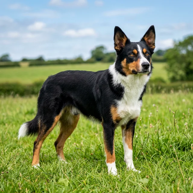
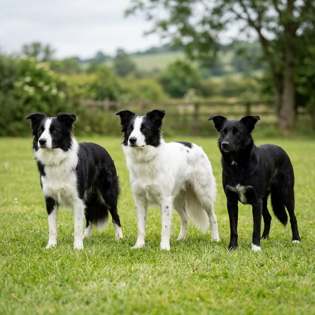
The classic Border Collie black and white is most often a/a (recessive black) or produced by KB. Tricolor (black, white, and tan) results when the K locus is ky/ky and the A locus is at/at or at/a.
Breeding note: Sable (Ay) is dominant over tan point (at), so a sable × tan point cross can produce sable puppies. Visual identification may be unreliable — DNA testing provides definitive answers.
B Locus (Brown / TYRP1) — The Chocolate Gene
Gene: TYRP1 (Tyrosinase-Related Protein 1)
The B locus determines eumelanin tone. The normal allele (B) produces black eumelanin, while the mutant (b) reduces TYRP1 enzyme function, expressing eumelanin as chocolate brown.
| Genotype | Eumelanin Color | Nose Color |
|---|---|---|
| B/B | Black | Black |
| B/b | Black (carrier) | Black |
| b/b | Chocolate / Brown | Liver (brown) |
Importantly, the B locus affects only eumelanin, not phaeomelanin. A b/b dog’s tan markings or sable reddish-brown areas remain essentially unchanged — only the black portions (and nose, eyelid rims, and pads) change color.
Multiple TYRP1 mutations (bs, bd, bc) have been reported in Border Collies, all producing the chocolate phenotype in homozygous or compound heterozygous states.
D Locus (Dilution / MLPH) — Color Dilution
Gene: MLPH (Melanophilin)
The D locus encodes melanophilin protein, which is involved in intracellular melanosome transport. The mutant allele (d) causes uneven melanosome distribution, creating a “washed out” dilution effect.
| Genotype | Effect | On Black Base | On Brown Base |
|---|---|---|---|
| D/D | Undiluted | Black | Chocolate |
| D/d | Undiluted (carrier) | Black | Chocolate |
| d/d | Diluted | Blue | Lilac (Isabella) |
Two dilution allele variants have been reported: d1 and d2. In Border Collies, d1 is primarily relevant, though in some breeds only d2 causes dilution. Comprehensive testing for both alleles is advisable in breeding programs.
Color Dilution Alopecia (CDA)
Dilute-colored individuals (blue, lilac) carry a risk of Color Dilution Alopecia (CDA) — a skin condition in which abnormal melanosome clumping damages hair follicles, causing progressive hair loss localized to dilute-colored areas.
Key points to emphasize:
- CDA does not develop in every d/d individual
- CDA onset involves modifier genes beyond the D locus
- With proper lineage management, many blue and lilac dogs live fully healthy lives
- The equation “dilute color = unhealthy” is scientifically inaccurate
At ROSCH KENNEL, breeding dogs with dilute coloring undergo thorough investigation of their lineage’s CDA history, and only lines with no CDA occurrence are used in breeding.
M Locus (Merle / PMEL17) — Beauty and Risk in One Gene
Gene: PMEL17 (also known as SILV/GP100)
The M locus is the most critical and most carefully managed locus in Border Collie coat color genetics. The merle gene results from a SINE (Short Interspersed Nuclear Element) insertion into the PMEL17 gene. Variations in the length of this SINE insertion (poly-A tail) produce multiple allele variants.

Merle Allele Variants
Recent research has revealed that the merle gene is not a simple M/m binary but rather a continuous spectrum based on SINE insertion length.
| Allele | SINE Length (bp) | Phenotype | Health Risk |
|---|---|---|---|
| m (non-merle) | — | Solid (no merle) | None |
| Mc (cryptic merle) | 200-230 | Appears solid, or very faint merle patches | Low |
| Mc+ (atypical merle) | 231-246 | Faint/irregular merle pattern | Low |
| Ma (atypical merle) | 247-254 | Incomplete merle pattern | Moderate |
| Ma+ | 255-264 | Near-merle pattern | Moderate to high |
| M (classic merle) | 265-268 | Typical merle pattern | High risk if M/M |
| Mh (harlequin merle) | 269+ | Merle with large white areas | High risk |
The Cryptic Merle Problem
Cryptic merle (Mc) is one of the most dangerous hidden risks. Dogs with this allele appear completely solid-colored (not visibly merle) while genetically carrying a merle allele.
If a cryptic merle is mistakenly identified as “solid” and bred with another merle, unexpected double merle puppies may result.
This risk is particularly acute in ee-red (e/e) individuals, where the merle pattern is poorly visible on phaeomelanin-based coat — sometimes called “phantom merle,” detectable only through DNA testing.
Double Merle (M/M) Health Risks — Why Merle-to-Merle Breeding Must Be Avoided
Double merle — individuals homozygous for the merle allele (M/M) — face severe health risks.

Hearing impairment: The merle gene affects melanocyte (pigment cell) development. The cochlea contains melanocytes essential for normal hearing. In double merles, these melanocytes are absent, causing degeneration of the stria vascularis and congenital sensorineural deafness — unilateral in some cases, complete bilateral deafness in others.
Visual impairment: Melanocytes are also involved in eye development. Double merles have been documented with:
- Microphthalmia — Underdeveloped eyeballs
- Iris coloboma — Partial absence of the iris
- Dyscoria/Corectopia — Pupil deformation or displacement
- Lens abnormalities — Positional anomalies or absence
- Retinal dysplasia — Structural retinal abnormalities
White coat area and disability correlation: Research demonstrates that larger white coat areas around the head correlate with higher risk of hearing and visual impairment, because melanocyte absence in the cranial region directly impacts sensory organ development.
Merle Breeding Rules
| Breeding Pair | Outcome | Risk |
|---|---|---|
| m/m × m/m | All puppies m/m (solid) | None |
| M/m × m/m | 50% M/m (merle), 50% m/m (solid) | None |
| M/m × M/m | 25% M/M (double merle), 50% M/m, 25% m/m | ⚠️ 25% double merle |
| M/M × m/m | All puppies M/m (merle) | None (parent is double merle) |
Rule: Merle × merle breeding must never be performed.
However, merle × solid breeding is entirely safe and produces beautifully patterned merle puppies without health risk. The merle gene itself is not dangerous — what’s dangerous is the homozygous state (M/M).
S Locus (White Spotting / MITF) — White Marking Extent
Gene: MITF (Microphthalmia-Associated Transcription Factor)
The S locus controls the extent of white markings (white spotting) on the coat. The characteristic Border Collie white blaze (white facial stripe), collar (white neck ruff), and socks (white paw tips) are all determined by this locus.
| Pattern | Description |
|---|---|
| Solid (S/S) | Nearly fully pigmented, minimal white |
| Irish Spotting | The classic Border Collie pattern. Blaze, collar, chest, socks |
| Piebald (sp/sp) | Large white areas. Body may be over 50% white |
| Extreme White | Nearly all white. Only small pigmented areas remain |
Excessive White and Hearing Impairment
As with the M locus, excessive white markings from the S locus also correlate with hearing impairment risk. The mechanism mirrors double merle: absence of melanocytes in the inner ear.
Individuals with fully white heads and ear regions show significantly elevated risk for congenital sensorineural deafness. In Border Collie breeding, managing white extent within appropriate bounds is essential.
Genotype-Phenotype Combinations — Common Colors and Their Genetics
The interplay of all seven loci produces the Border Collie’s remarkable coat color diversity. Representative color patterns and their genotypes:
| Color | E | K | A | B | D | M | Nose |
|---|---|---|---|---|---|---|---|
| Black & White | E/- | ky/ky | a/a | B/- | D/- | m/m | Black |
| Black Tricolor | E/- | ky/ky | at/at or at/a | B/- | D/- | m/m | Black |
| Chocolate & White | E/- | ky/ky | a/a | b/b | D/- | m/m | Liver |
| Chocolate Tricolor | E/- | ky/ky | at/- | b/b | D/- | m/m | Liver |
| Blue & White | E/- | ky/ky | a/a | B/- | d/d | m/m | Slate gray |
| Lilac & White | E/- | ky/ky | a/a | b/b | d/d | m/m | Pinkish brown |
| Blue Merle | E/- | ky/ky | a/a | B/- | D/- | M/m | Black (partial pink) |
| Red Merle | E/- | ky/ky | a/a | b/b | D/- | M/m | Liver |
| Slate Merle | E/- | ky/ky | a/a | B/- | d/d | M/m | Slate gray |
| ee-Red | e/e | — | — | B/- | D/- | m/m | Black |
| Sable | E/- | ky/ky | Ay/- | B/- | D/- | m/m | Black |
※ ”—” indicates any dominant allele or any allele.
Breeding Simulations — Practical Case Studies
Case 1: Black & White × Chocolate & White
Sire: Black & White (B/b, D/D, a/a, m/m) Dam: Chocolate & White (b/b, D/d, a/a, m/m)
B locus Punnett Square:
| b | b | |
|---|---|---|
| B | B/b | B/b |
| b | b/b | b/b |
→ 50% Black (B/b), 50% Chocolate (b/b)
D locus:
| D | d | |
|---|---|---|
| D | D/D | D/d |
| D | D/D | D/d |
→ 50% D/D, 50% D/d, 0% d/d — No dilute puppies will be produced
Expected puppies: Black & White (50%), Chocolate & White (50%). All puppies carry DD or Dd. Future Dd × Dd matings could produce blue or lilac offspring.
Case 2: Blue Merle × Black & White (Safe Breeding)
Sire: Blue Merle (B/B, D/D, a/a, M/m) Dam: Black & White (B/b, D/d, a/a, m/m)
M locus:
| m | m | |
|---|---|---|
| M | M/m | M/m |
| m | m/m | m/m |
→ 50% Merle (M/m), 50% Solid (m/m) → Double merle risk: Zero ✅
Factoring in other loci, this cross may produce blue merle, black & white, and possibly chocolate merle (if B/b) puppies.
Case 3: ⚠️ Merle × Merle (Prohibited Breeding)
Sire: Blue Merle (M/m) Dam: Red Merle (M/m)
| M | m | |
|---|---|---|
| M | M/M | M/m |
| m | M/m | m/m |
→ 25% Double Merle (M/M) ⚠️ High risk of hearing and visual impairment → This breeding must never be performed
Dispelling Common Misconceptions About Coat Color Genetics
”Dilute Colors Are Prone to Disease” — Half Right, Half Wrong
It’s true that blue and lilac dogs carry CDA risk. But not every dilute-colored dog develops CDA. Onset is heavily influenced by modifier genes and varies substantially by family line.
Blue and lilac Border Collies from well-managed bloodlines are often just as healthy and long-lived as their non-dilute counterparts. Judging a dog’s health by color alone is scientifically unsound.
”Merle Is a Dangerous Color” — Accurate Understanding Required
The merle gene itself poses no danger. Heterozygous (M/m) merle dogs carry no elevated health risks compared to non-merle dogs. The danger lies exclusively in homozygous (M/M) double merle — which is 100% preventable by never breeding merle to merle.
Categorically condemning merle as “dangerous” is as misguided as it is unnecessary. What matters is managing it with proper genetic knowledge.
”Confusing Ticking with Merle”
Ticking (T locus) produces small pigmented spots scattered through white areas. At a distance, it can resemble blue merle, but the mechanisms are entirely different.
- Merle: Random dilution of pigmented areas, creating patches of varying intensity. Operates at the melanosome level.
- Ticking: Small pigmented dots appearing in white areas. Caused by secondary migration of pigment cells.
Cases of inexperienced breeders misidentifying heavily ticked dogs as “merle” — or vice versa — have been documented. DNA testing provides the definitive answer.
The “Tweed” Pattern Explained
“Tweed” describes a merle phenotype with large patches of different color intensities within the merle areas. This is not caused by a separate gene but rather by somatic reversion of the merle allele — mutations in the SINE poly-A tail length occurring during cell division, producing a mosaic of different merle expressions.
DNA Testing in Practice — Utilizing Coat Color Gene Panels
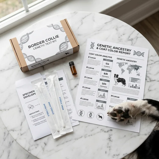
Coat color DNA testing is now easily performed through buccal (cheek) swab collection. ROSCH KENNEL tests all breeding dogs for the following loci, primarily using Orivet’s color panel:
- E locus (MC1R)
- K locus (CBD103)
- A locus (ASIP)
- B locus (TYRP1 — bs, bd, bc mutations)
- D locus (MLPH — d1, d2 mutations)
- M locus (PMEL17 — quantitative SINE insertion length)
- S locus (MITF)
All results for every breeding dog are 100% publicly available. Transparency is the bedrock of trust and the cornerstone of science-based breeding.
Frequently Asked Questions (FAQ)
Q: I’d love a merle puppy, but I’m worried about health.
A: A merle (M/m) dog is just as healthy as a non-merle dog. The concern is exclusively with double merle (M/M), which is entirely preventable through merle × solid breeding only. All merle dogs at ROSCH KENNEL are DNA-tested, and merle-to-merle breeding is never performed. Rest assured.
Q: I’ve heard blue Border Collies are prone to CDA (a skin condition). Is this true?
A: CDA risk does exist, but it does not affect every blue dog. Onset is heavily influenced by genetic lineage. ROSCH KENNEL breeds dilute colors exclusively from lines with no CDA history, minimizing risk.
Q: Can you predict puppy coat colors from the parents?
A: With DNA test results establishing both parents’ genotypes, puppy color variations can be predicted with high accuracy. However, which color any individual puppy will express remains probabilistic. That’s part of nature’s richness.
Q: Is it true that you can’t visually determine whether an ee-red (golden red) dog is merle?
A: Correct. Since ee-red dogs produce only phaeomelanin in their coat, the merle pattern is poorly visible. DNA testing is the only reliable method, and M locus testing is mandatory before using any ee-red dog in a breeding program.
Q: What are your thoughts on “rare colors” being sold at premium prices?
A: Placing excessive premiums on color rarity incentivizes breeding that prioritizes appearance over health. Rare colors like lilac merle or blue tricolor are beautiful, but they should never be the goal driving breeding decisions. Color is a result, not an objective. The priority is producing healthy dogs with stable temperament — colors that emerge naturally from well-planned genotype combinations are something to be appreciated, not engineered at the expense of wellbeing.
ROSCH KENNEL’s Breeding Philosophy — If It Can Be Measured, We Measure It
- Full genetic testing on all breeding dogs — 15+ hereditary disease markers + 7 coat color loci (this is the minimum standard)
- Merle × merle breeding is never performed — Zero double merle risk
- Carrier × carrier breeding is never performed — Eliminating known hereditary disease onset risk
- 100% public disclosure of all test results — Transparency is the foundation of trust
- Coefficient of Inbreeding (COI) management — Maintaining genetic diversity and avoiding inbreeding
- Dilute colors bred exclusively from CDA-free lineages — Decisions based on family history data
- HD/ED (joint) palpation assessment on all dogs — Addressing multifactorial conditions not covered by DNA testing
- Ongoing research and observation of conditions beyond current test capabilities — Tests are a minimum benchmark, not the final word
Our breeding program anchors on black as the primary axis while implementing rigorously data-driven pairing decisions. This approach produces beautiful, healthy dogs with consistency that spans generations.
Closing Thoughts — Science Illuminates, Breeder Awareness Protects
Border Collie coat color genetics is deep, fascinating, and occasionally entails complex health considerations. That’s precisely why breeding must be guided by the combined power of science, data, and experience — not mere pursuit of visual appeal.
DNA testing is a powerful tool for understanding coat color genetics, but it does not guarantee a dog’s overall health. Testing is the baseline — a minimum benchmark. What lies beyond it — the breeder’s daily observation, the quality of the rearing environment, the commitment to in-person placement — is what truly sustains healthy dogs.
Color is part of a dog’s appeal, but it is never the purpose of breeding. Genotype, health risk, temperament, and the rearing environment have to be handled within the same plan if the next generation is to be protected.
References & Resources
- Langevin, M. et al. (2018). “Merle phenotypes in dogs — SILV SINE insertions from Mc to Mh.” PLOS Genetics, 14(2).
- Clark, L.A. et al. (2006). “Retrotransposon insertion in SILV is responsible for merle patterning of the domestic dog.” Proceedings of the National Academy of Sciences, 103(5), 1376-1381.
- Strain, G.M. (2004). “Deafness prevalence and pigmentation and gender associations in dog breeds at risk.” The Veterinary Journal, 167(1), 23-32.
- Schmutz, S.M. & Berryere, T.G. (2007). “Genes affecting coat colour and pattern in domestic dogs: a review.” Animal Genetics, 38(6), 539-549.
- Everts, R.E. et al. (2000). “Identification of a premature stop codon in the melanocyte-stimulating hormone receptor gene (MC1R) in Labrador and Golden retrievers with yellow coat colour.” Animal Genetics, 31(3), 194-199.
- Philipp, U. et al. (2005). “Polymorphisms within the canine MLPH gene are associated with dilute coat color in dogs.” BMC Genetics, 6, 34.
Daily observation is where the next article begins.
Instagram shows the dogs in daily life. The journal turns the themes we notice there into evidence-aware resources.